Melanoma Pictures
Here are the greatest melanoma images for you to download in order to recognize any possible symptoms of the disease on your own body. You might need to see a doctor if the symptoms in the image closely match yours. Making a picture comparison and consulting your doctor about this issue right away is, thus, highly appropriate. For those who are interested in learning more about the impacts of melanoma on the human body, we are providing downloadable photographs. The melanoma kind was used to group the photos. Using these photos, you can establish if you have melanoma. Download the large-size photos to your computer if you want to see a crisper image. You can then enlarge the image to see the mole in greater detail. In order to identify the disease symptoms, you can compare these moles with your physical symptoms. the consequences of melanoma illness. Prior to the pleural effusion, hydrothorax is a feature of melanoma. Fluid accumulation in the lungs is known as a pleural effusion. But if I may, I should highlight the moderate to severe discomfort on the sides of the body as a symptom of this problem.
The network of nerves in the pleura, which is so delicate, is extremely dense. There are two thin membranes, one of which covers the lung’s surface and the other of which covers the inside of the chest. In the chest, these two membranes store 300–500 cm3 of fluid and secrete certain substances. Numerous elements are absorbed and released by the body through this system. This creates an oleaginous surface in the chest that allows the lungs to move when breathing. Through the pleura, a healthy human body transfers water often. However, a sick body with lung conditions excretes the water but has trouble absorbing it. Here, extra bodily water collects and puts pressure on the area, causing pain. The lungs should be CT examined even if only a little alteration is found on the pleura.
Describe melanoma
Melanoma is a cancer that affects people all over the world, with an increasing prevalence and a difficult treatment regimen. If the disease is discovered in its early stages and affects the top layer of skin, the chance of recovery will be higher.
But if the melanoma has spread throughout the body, the chances of recovery are drastically reduced or even eliminated. However, a novel therapy known as CICD is found in the UK in 2017 and appears to hold the promise of curing skin cancer. However, there is currently no medication that can completely cure melanoma.
Facts about melanoma
- One of the top 10 factors leading to new cancer cases is melanoma.
- Melanoma is a rare type of cancer that has the capacity to spread significantly to other areas of the body.
- Melanomas can develop in locations that are not exposed to sunlight.
- People under the age of 20 are diagnosed with 2% of all melanoma cases.
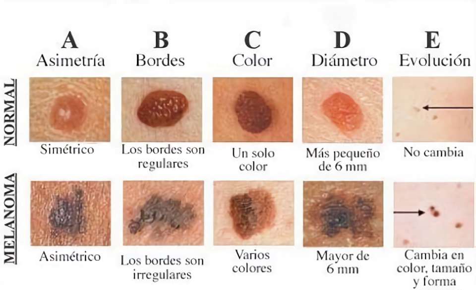
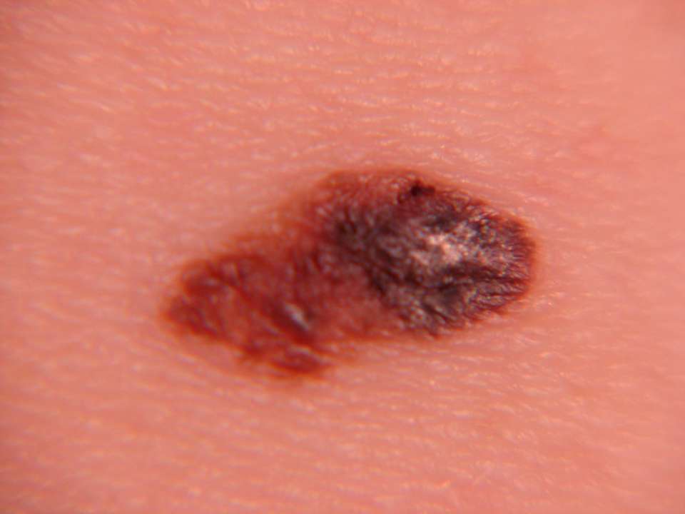
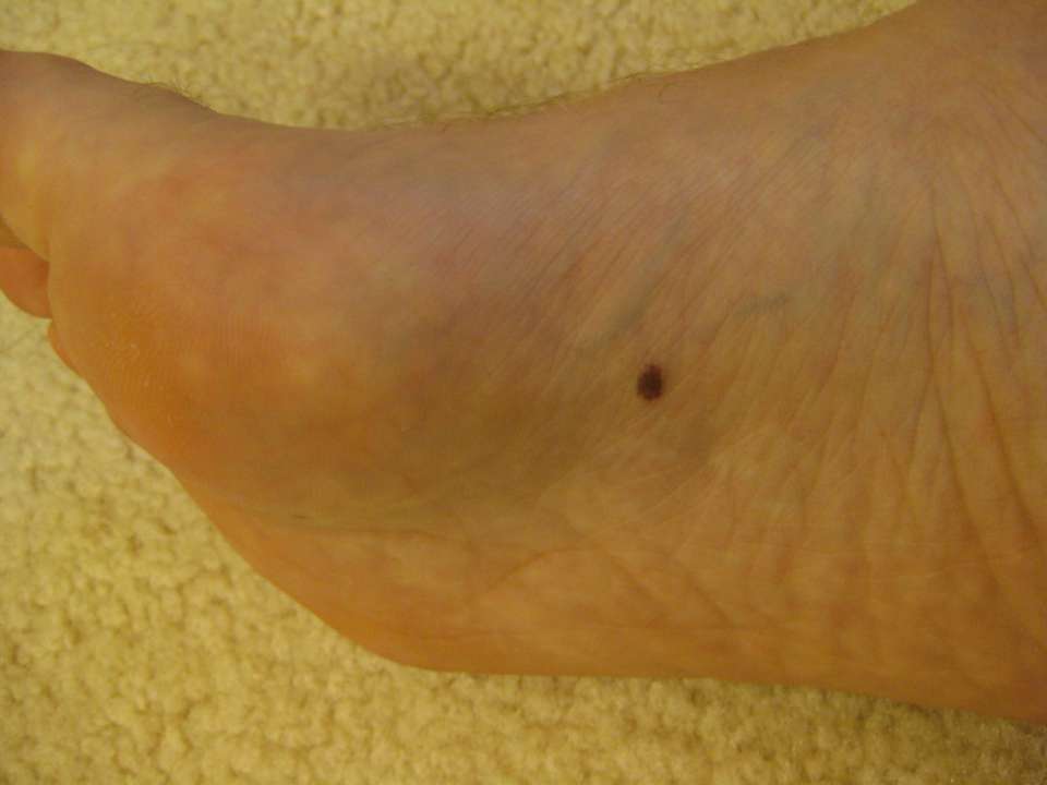
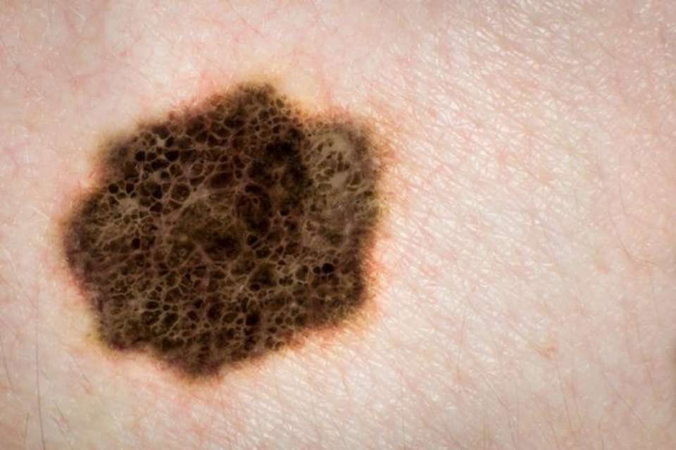
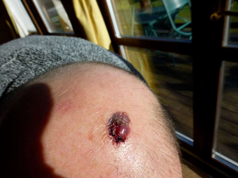
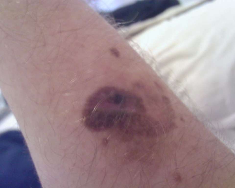
Symptoms of Melanoma in Pictures

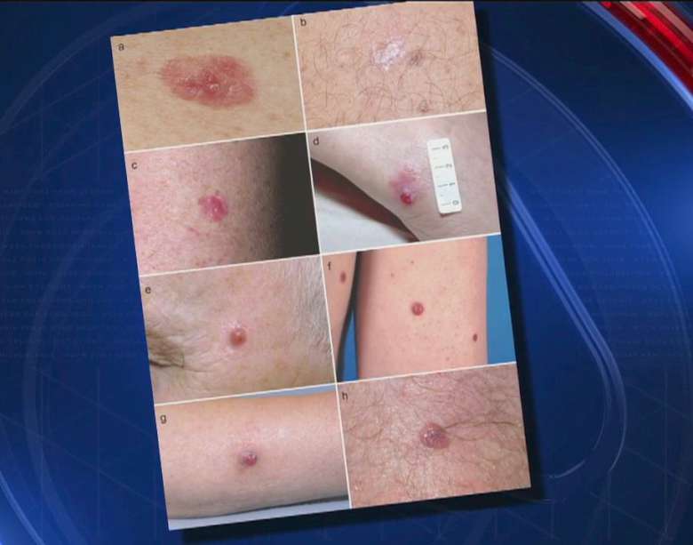
Is it feasible to recognize melanoma from a picture of the symptom alone? Actually, a body’s visual examination alone cannot conclusively rule out skin cancer. However, you can get a general understanding by contrasting the pictures we offer with the unsettling skin signs. So that you can aid in the early discovery of the sickness, you can see a doctor.
It is quite beneficial for this crucial step to compare the moles on your skin with the melanoma moles depicted in these photographs. Consult your doctor right away if you notice any moles on your skin that are similar. Early detection of melanoma moles gives you the chance to visit a hospital before it’s too late and receive an effective treatment.
If the fluid is found by a CT scan and the patient’s complaints match, the diagnosis will be determined. There are two basic categories in which to classify pleura disorders. The first one is an inflammatory pleural effusion, and the second one is brought on by a tumor. These are actually dry and moist inflammation. Thoracentesis, or the removal of fluid from the pleura, should be performed in the event of even a little irritation or thickening.
The medical procedure
It is important to test the fluid analysis for both common microorganisms and Mycobacterium tuberculosis. Since the mycobacterium TB is the microorganism that affects the pleura most frequently, it should undoubtedly be looked at. When the lungs are inflamed and thick, tuberculosis is a strong candidate as the cause. After checking for tuberculosis, it is also important to take into account the other diseases since they can also cause these symptoms.
If inflammatory cells are found in the fluid that was extracted, the patient is typically given antibiotics and other drugs, and the follow-up procedure is then continued. If the fluid begins to drain from the lungs in one or two months, the doctors will temporarily halt the medication and monitor the condition.
Sometimes, this medication may thicken the lungs, and it may even erode immunological defenses. Surgery is used to remove the pleura if medicine fails to reduce pleural thickness since the lungs will continue to function normally after the procedure.
If after the analysis tuberculosis is found, the patient is given medicine for this illness for at least three months. Alternatively, the maximum waiting period is three months if the inflammation and tuberculosis appear to remain stable on the CT scan after three months. The phthisic pleura is surgically cleared after three months, and the lungs resume normal function.
Complaints
Chest pain and breathing problems are the most frequent complaints in pleural carcinoma cases. Additionally possible symptoms include exhaustion, anemia, coughing up blood (hemoptysis), and fatigue. Inflammation on the neck and face, as well as difficulties swallowing, may be seen if the cancer has migrated to tissues outside of the pleura.
Findings
Melanoma is not specifically found during an examination. This condition may also exhibit the typical symptoms of any lung disease, such as a reduction in breathing movements and sounds on the side of the affected lung.
How do you diagnose?
Advanced diagnostic techniques should be used to assess a patient with a history of mesothelioma and inflammation or thickness on the pleura seen on chest radiographs and CT scans. A pleural sample is necessary for the accurate diagnosis even though chest radiography and CT scans also reveal the typical abnormalities.
The fluid sample (specimen) obtained from the lung is initially analyzed for a pathologic report. A second pleura specimen is obtained via pleural biopsy if this report is insufficient to make a final diagnosis. Either a surgical biopsy or a needle biopsy can be used to get the specimen. To track the development of cancer, more sophisticated techniques like PET-CT, ultrasonography, and MR (Magnetic resonance) may also be used.