Melanoma Pictures Free Download
When it comes to understanding melanoma, visual resources can be incredibly valuable. That’s why I’m excited to share a selection of images that showcase the various appearances of this skin cancer. By examining these pictures, you can get a clearer idea of how melanoma manifests at different stages. As many know, early detection is vital—if you observe any changes to your skin, seeking a professional’s assessment can be life-saving. These images serve as an educational guide to help you recognize melanoma and identify warning signs.
Let’s dive into the images that highlight the many facets of melanoma.
Understanding Melanoma on the Skin
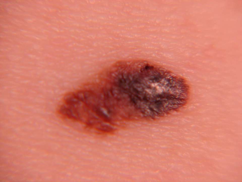
One striking photo captures the typical features of melanoma. In this image, you’ll notice a spot characterized by irregular borders, a variety of dark colors, and an asymmetric shape. These distinct visual traits are clinical indicators of melanoma and warrant immediate medical evaluation. Since melanoma treatment is most effective when caught early, it’s critical to have anything suspicious examined by a dermatologist as soon as possible.

Melanoma vs. Normal Moles
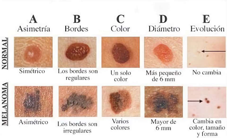
Another important illustration lays out the key distinctions between melanoma and typical moles using the ABCDE method. This established criteria includes:
- A (Asymmetry): Normal moles tend to be symmetrical, while melanoma often presents an asymmetric shape.
- B (Borders): A typical mole boasts smooth, even edges. In contrast, melanoma often has irregular, ragged borders.
- C (Color): Normal moles are usually a single shade, whereas melanoma displays multiple colors.
- D (Diameter): Moles are generally smaller than 6mm, while melanoma can be larger.
- E (Evolution): Normal moles maintain a consistent appearance over time, but melanoma can swiftly change in size, shape, or color.
This framework is crucial for early detection and reminds us all to monitor any skin changes closely and consult a dermatologist as needed.
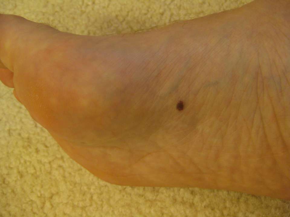
Melanoma Under the Feet
In another striking visual, a melanoma lesion is captured on the sole of the foot. Against lighter skin, a dark brown spot stands out, relatively small and round, lacking obvious abnormalities around it. If you notice something like this, it is important to see a doctor immediately.
Melanoma on the Body
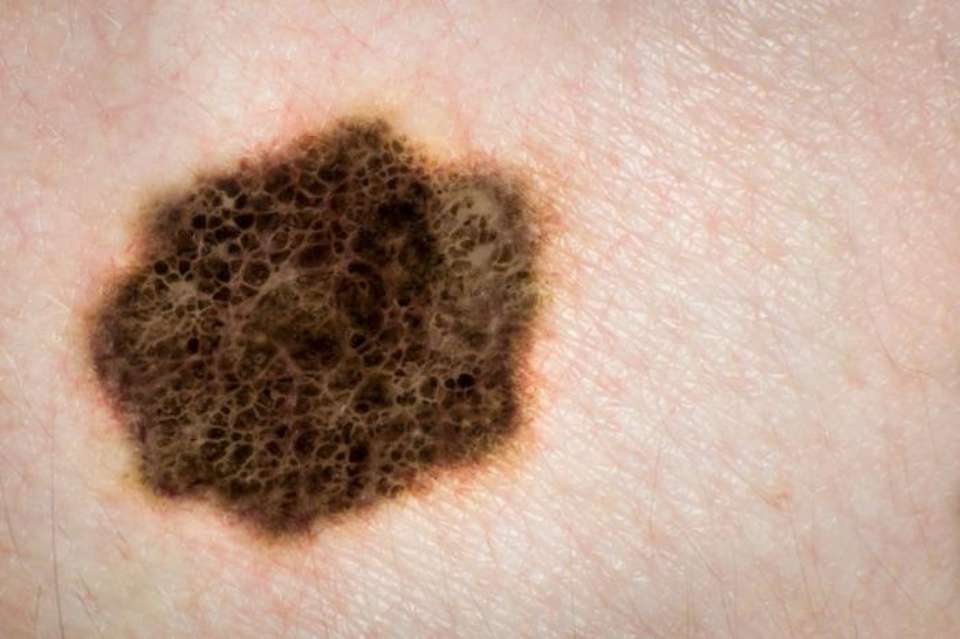
One image focuses on an irregular dark spot located on light skin. The jagged borders and uneven color, with shades of black, dark brown, and lighter brown blending unpredictably, are concerning features. There’s even a net-like pattern within the lesion that could indicate melanoma, underlining the need for timely consultation with a specialist.
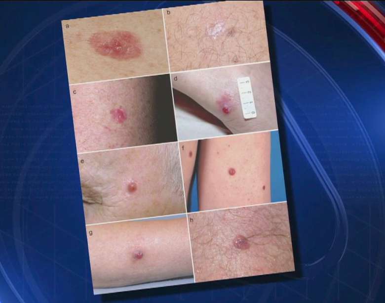
Melanoma on the Knee
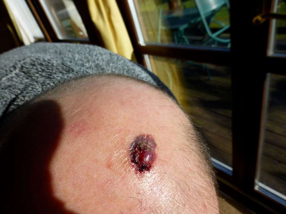
Another photo depicts a reddish-purple, raised lesion on a knee. The surrounding skin appears slightly inflamed, and the lesion seems firm and swollen, perhaps even scabbed over. With irregular borders and noticeable color changes, this type of find could suggest a more advanced stage of skin cancer, again necessitating urgent medical attention.
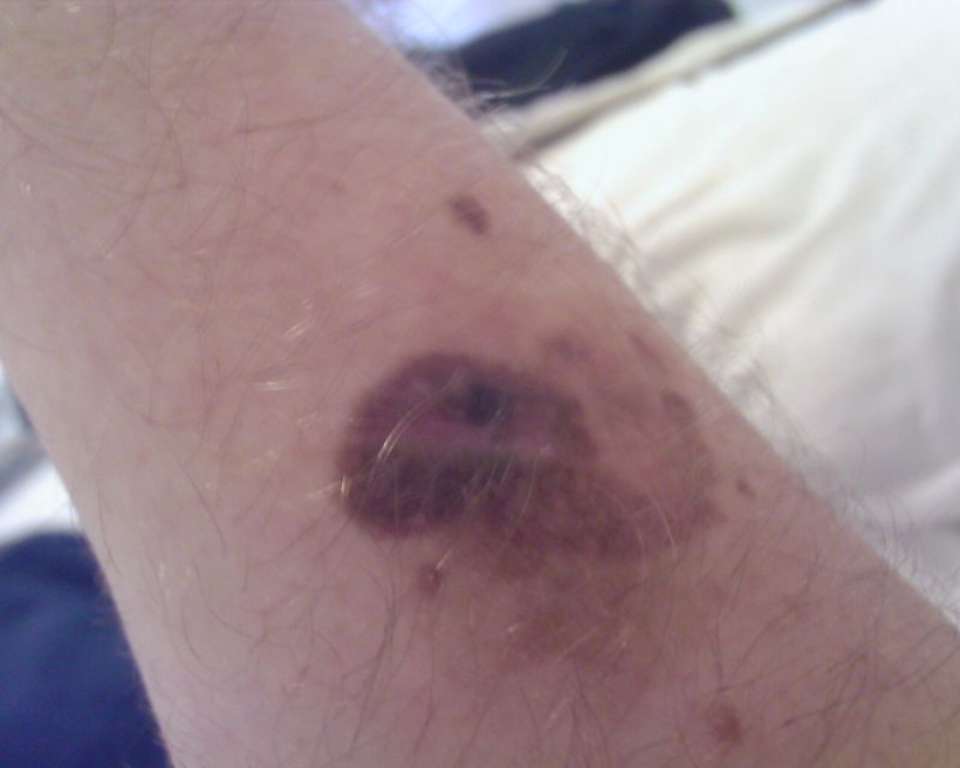
Melanoma on the Arm
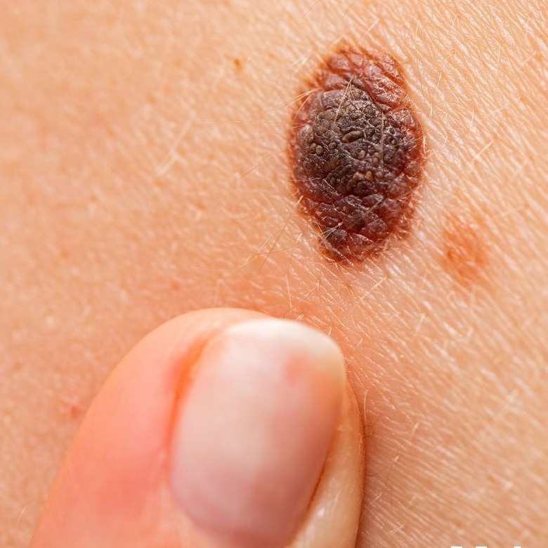
A closer look at an irregular dark brown spot on an upper arm demonstrates uneven borders and varying shades, with lighter areas at the edges and a darker center. Nearby smaller brown spots add to the complexity of this lesion. Given these irregular characteristics, a professional evaluation from a dermatologist is essential.
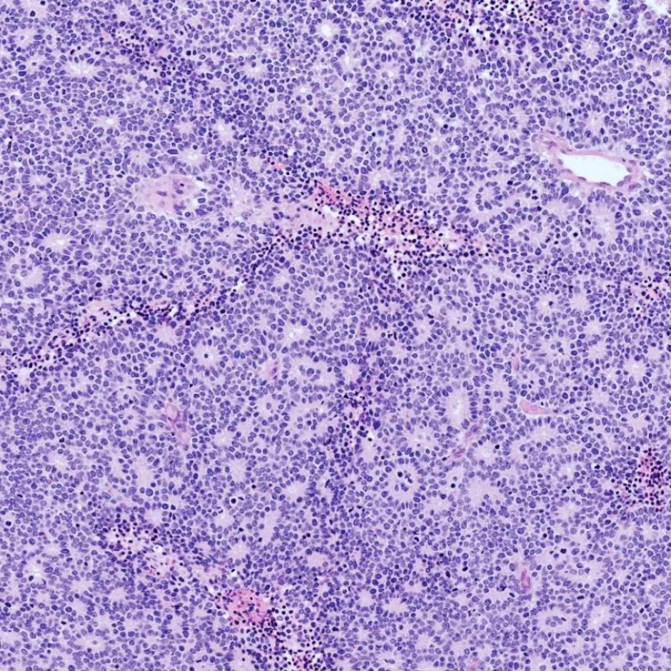
Retinoblastoma Under the Microscope
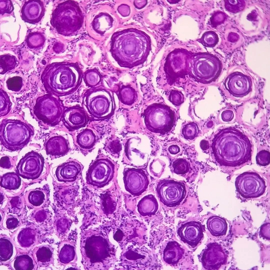
Switching gears, we take a look at a microscopic image of retinoblastoma, a different type of cancer. This stained tissue shows the malignant cells grouped in circular patterns, with lighter areas suggesting blood vessels in between. This kind of analysis is typically performed by pathologists and is crucial for understanding the nature of tumors.
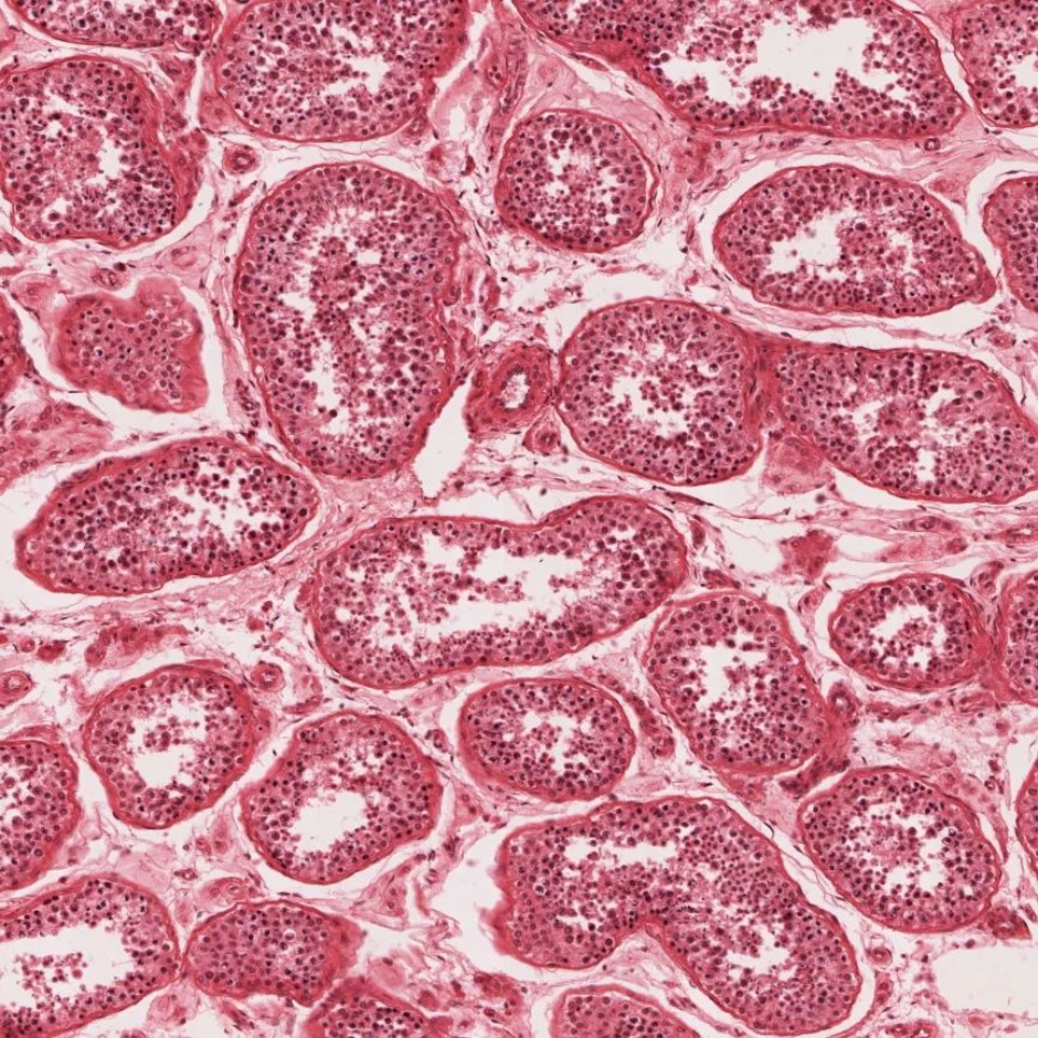
Spermatogenesis in the Testis
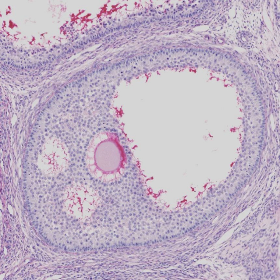
Lastly, an enlightening microscopic image depicts spermatogenesis in testis tissues. Labeled sections show different stages involved in sperm production:
- Spermatogonia: These stem cells located in the outer layer are essential for initiating sperm production.
- Primary Spermatocytes: Larger cells that arise after spermatogonia division, setting the stage for meiosis.
- Spermatids: These round cells result from meiosis and begin transforming into sperm.
- Smooth Muscle: Surrounding the tubules, this muscle layer supports fluid movement and cell dispersion.
This well-organized structure is a staple in education regarding reproductive biology and histology.
Understanding melanoma is crucial for proactive health management. These images serve not only as informative tools but also as a reminder of the importance of skin health. If you have questions or concerns regarding moles or skin changes, don’t hesitate to reach out to a healthcare provider. Early action can lead to successful treatment and peace of mind.
Understanding Melanoma and Its Diagnosis: What You Need to Know
When it comes to skin health, melanoma stands out as one of the most serious forms of skin cancer. Its rising prevalence in recent years makes awareness crucial for everyone. Early diagnosis can be a game-changer, offering a better chance for successful treatment. Ignoring the signs can lead to advanced stages of the disease, which are much harder to tackle.
First things first, what exactly is melanoma?
It’s a type of skin cancer that can develop not only on areas exposed to sunlight but also in places where the sun rarely shines. It’s important to keep an eye on moles — their size, shape, and color can tell you a lot about potential issues. The use of sunscreen daily and limiting sun exposure are essential steps for protecting your skin against this formidable cancer.
Here are some key facts
Melanoma is now one of the most frequently diagnosed cancers around the globe. Surprisingly, it can affect anyone, regardless of skin type, although fair-skinned individuals are at a higher risk. Alarmingly, about 2% of melanoma cases occur in people under 20 years old. Regular self-exams and visits to a dermatologist can aid in early detection, ensuring that any signs are caught promptly.
So, what should you look for?
The hallmark of melanoma is a change in an existing mole—watch for irregular borders, changes in color, or rapid growth. Additional signs include open sores that crust or bleed. In some cases, melanoma may cause swollen lymph nodes as it spreads to other parts of the body, including crucial organs like the liver or lungs.
Dermatologists often utilize tools like dermatoscopy to get a closer look at suspicious moles. Catching melanoma early can significantly boost the chances of effective treatment. If you notice any unusual changes on your skin, don’t hesitate to consult with a dermatologist. Your skin is your body’s largest organ—let’s keep it healthy!
Understanding Pleural Issues and the Impact of Melanoma on Lung Health
If you’ve ever experienced a tightness in your chest or breathlessness, you might be dealing with a problem related to the pleura. This thin, delicate membrane surrounds your lungs, enabling them to expand and contract smoothly as you breathe. In a healthy body, the pleura produces and absorbs a small amount of fluid, but health issues can cause this fluid to build up, leading to uncomfortable and sometimes serious complications.
Symptoms of pleural problems can include difficulty breathing, chest pain, and discomfort in the back. To get to the bottom of these issues, medical professionals often rely on fluid analysis and advanced imaging techniques. An accurate diagnosis is essential to determine the right course of treatment.
One serious condition linked to pleural issues is pleural effusion, which can occur in advanced melanoma cases. Here, fluid accumulates around the lungs, causing symptoms like chest pain, shortness of breath, and excessive fatigue. To identify pleural effusion, doctors typically use CT scans to detect fluid buildup and analyze it for further insights.
The presence of pleural effusions can significantly affect a patient’s quality of life and complicate cancer treatment. Managing this condition often requires surgical intervention or the drainage of excess fluid. Regular checkups and preventative care are crucial in monitoring disease progression and ensuring the best possible outcomes.
When it comes to diagnosing melanoma, dermatologists typically start with a visual examination of the skin for any concerning changes. If they spot something unusual, a biopsy is performed to confirm their suspicions. For pleural complications, thorough fluid analysis and additional tests help rule out potential infections, including tuberculosis.
Treatment for melanoma depends on various factors, including the stage of cancer and the overall health of the patient. Options may include surgery and immunotherapy. Regular monitoring and personalized treatment plans play a significant role in managing the disease.
Early detection is key in managing both melanoma and pleural complications effectively. Don’t ignore mild symptoms; consulting with a healthcare specialist can protect your health.
If you notice any alarming changes in your skin—like a mole that grows quickly, alters in color, or has irregular borders—don’t hesitate to reach out to a dermatologist. Detecting melanoma early can be life-saving.
Experts consistently emphasize the importance of regular skin checks and adopting preventive measures against skin cancer. Simple habits like using sunscreen, avoiding excessive sun exposure, and staying vigilant about skin changes can significantly reduce your melanoma risk.
Stay on top of your health. Be aware of symptoms like chest pain, persistent fatigue, and abnormal moles, and consult a specialist promptly. Expert care can make all the difference; timely action is the key to a healthier, happier life.