Your guide to understanding that spot on your skin: Melanoma vs Normal moles (a visual guide).
I’ve put together a guide to help you make sense of that spot you’re wondering about. It includes free, downloadable comparison images showing side-by-side examples of melanomas and harmless (benign) moles that you can save for reference.
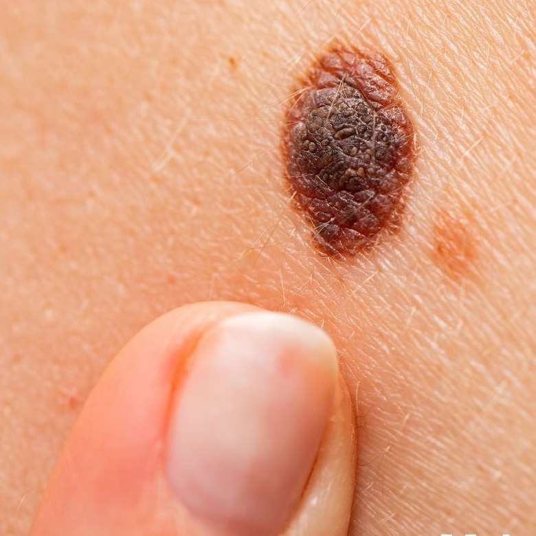
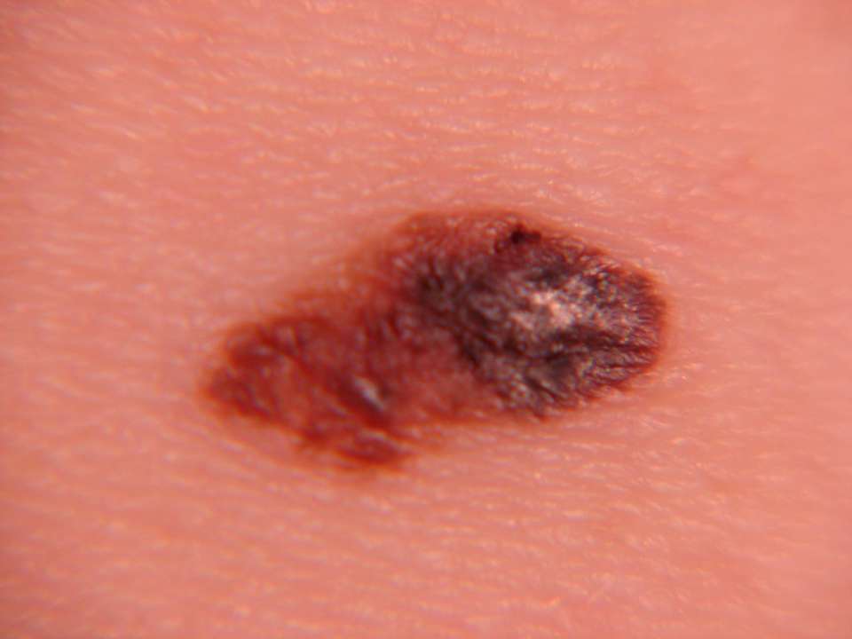
If you have a spot that makes you ask, “What is this?” take a moment to look at the detailed, close-up melanoma photos I’ve included below. You’ll be able to compare them to your own mole. If you notice a similarity and feel a hint of suspicion, don’t leave it to chance—schedule an appointment with a dermatologist right away. That small step could be one of the most important things you ever do for yourself.
But don’t panic just yet! Not every unusual mole means melanoma. My goal here isn’t to scare you, but to empower you. With the right information, you can let go of unnecessary worry and learn what truly deserves your attention. So let’s take a look together.
When looking for melanoma, remember these 3 golden rules:
Is the color uniform or mixed?
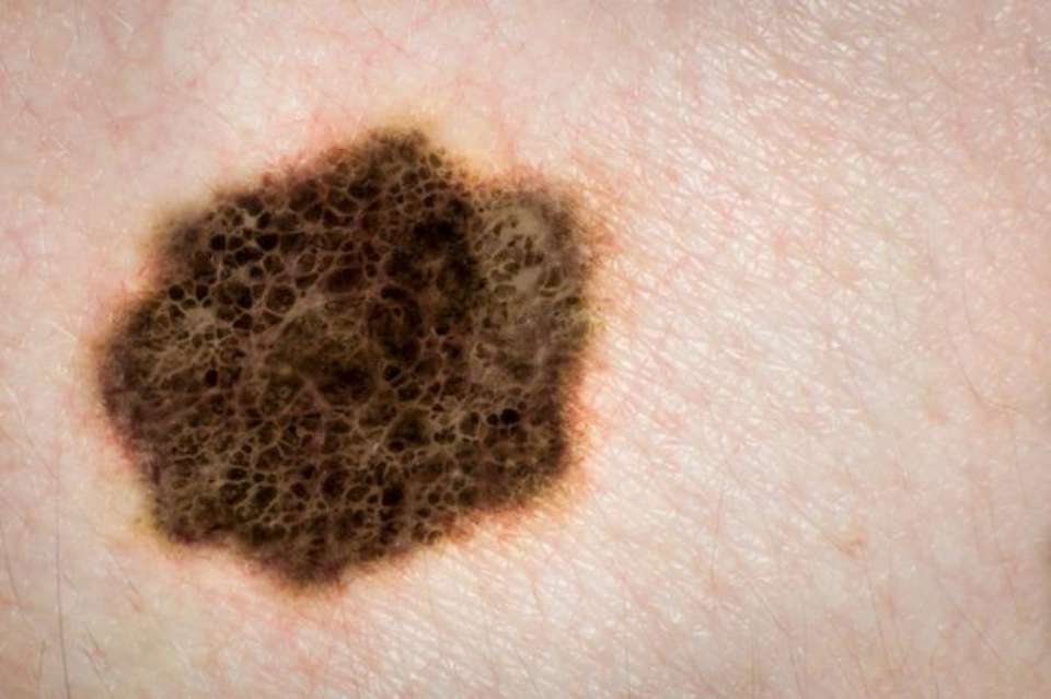
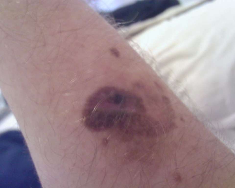
The first and most critical clue is color. Harmless moles are usually a single, uniform shade (often a shade of brown). With melanoma, things get complicated. Upon close inspection, you might see light brown, dark brown, even black, reddish or whitish areas all mixed together. If the palette has multiple colors, consider that your first alarm bell.
Are the borders sharp or blurry?
The second key point is shape and borders. Benign moles are typically smooth, round or oval with sharp, well-defined borders. Melanomas often have irregular, ragged, notched or “geographic” borders. The edges can appear blurry, as if the pigment is “leaking” into the surrounding skin.
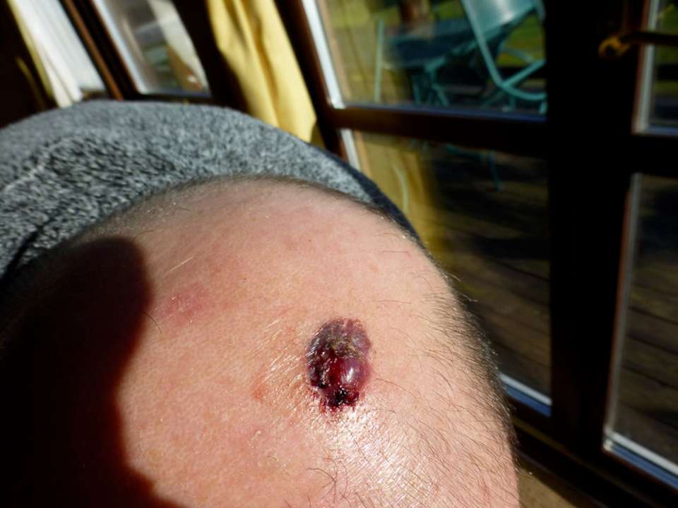
Change and behavior: is it growing? Bleeding?
The third and perhaps most personal, criterion is change. A benign mole usually stays the same for years. It doesn’t grow, bleed, itch or crust over. Melanoma is dynamic. It can change size, shape or color over months or even weeks. It might itch, bleed or form a crust. A general rule: “A spot you’ve had forever that never changes” is usually okay. “A new spot that appears and changes quickly” demands attention.
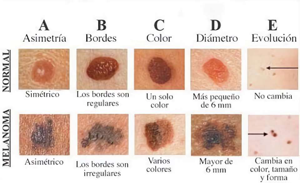
Comparison images: train your eye.
I’ve included a comparison board below showing normal moles and melanomas side-by-side. You’ll see that the top row features spots that are symmetrical, with smooth borders and uniform color. The bottom row shows spots that are irregular, multi-colored and asymmetrical. Use these visuals to train your eye.
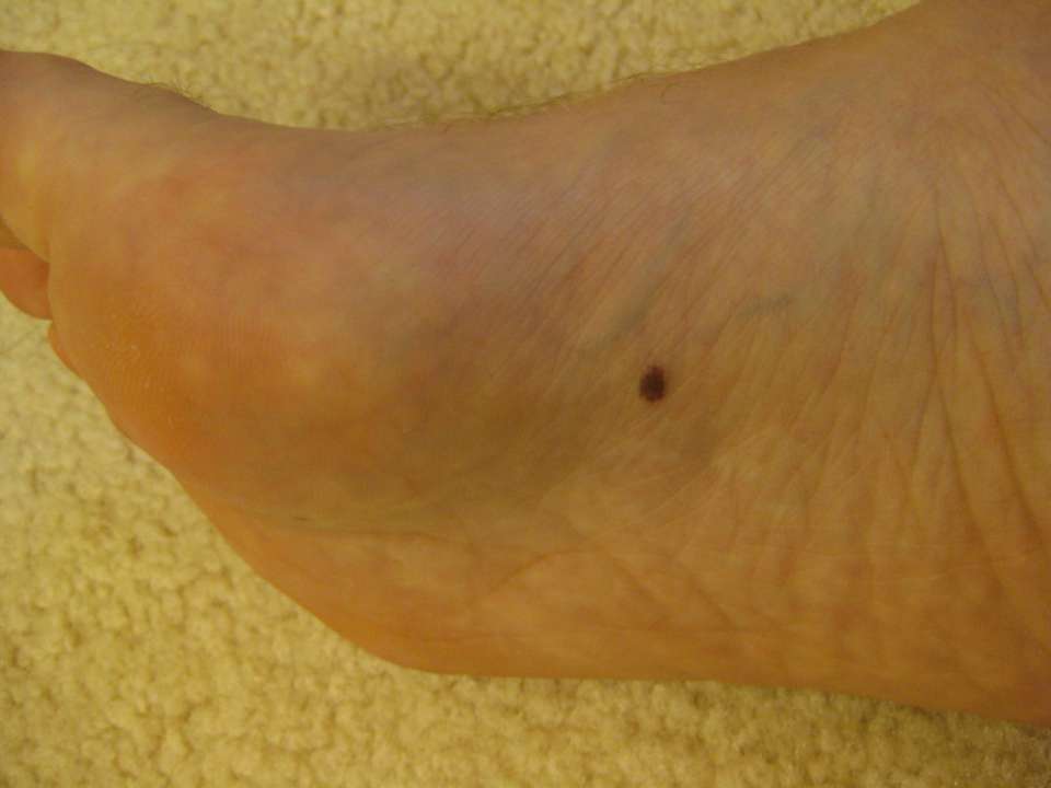
Special areas like soles and palms:
Small, dark spots on the soles of your feet or palms of your hands can be tricky. The most important question here is: “Is this spot new?” If you’ve had it since birth and it hasn’t changed, it’s probably nothing to worry about. But if it’s new or growing, you must get it checked. Remember: melanoma itself usually doesn’t hurt; pain is often a sign of another issue (like a bleed).
If you see a suspicious spot:
Please see a dermatologist promptly if your spot has:
- Multiple colors.
- Irregular, ragged borders.
- Rapid change in size, or if it’s bleeding, itching, or crusting.
Early detection is your greatest weapon.
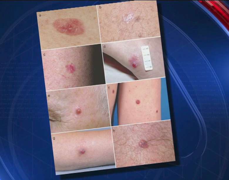
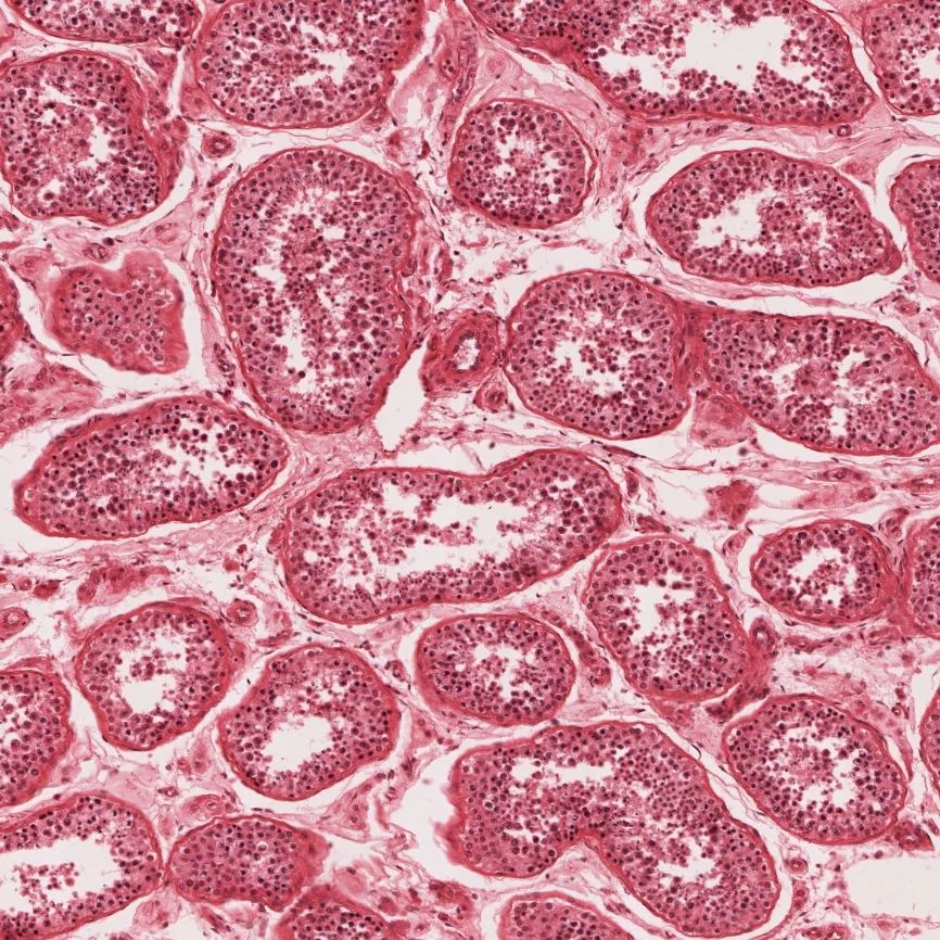
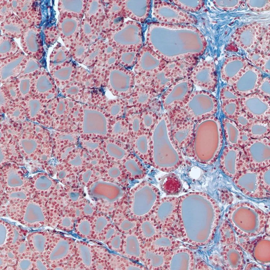
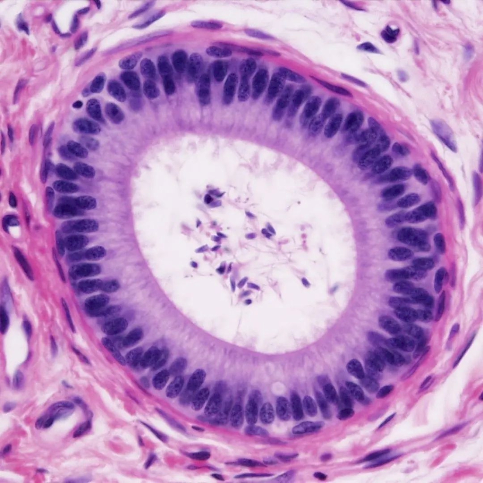
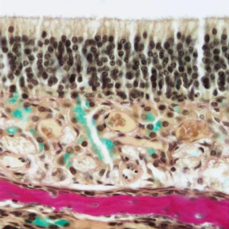
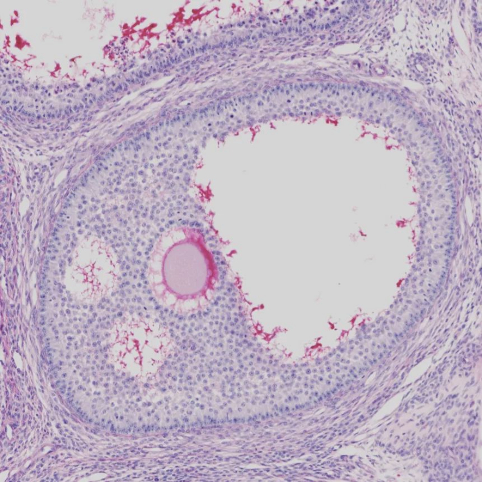
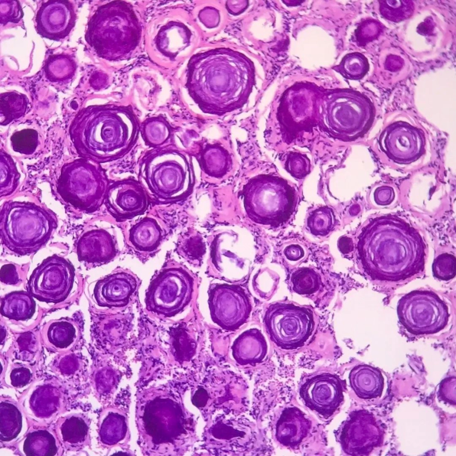
Melanoma under the microscope:
I’ve also included microscopic images of melanoma. These are to show how complex this subject is—not for you to try and self-diagnose. Use them to understand what your doctor is looking for. Leave the actual diagnosis to the trained professionals—dermatologists and pathologists—who know how to read these images.
I hope this information and these visuals help clear things up for you. When in doubt, the best thing to do is always to show it to a professional. Take good care of yourself!
PS: Feel free to use the download buttons to save the images or share this guide with a loved one.


Leave a Reply
You must be logged in to post a comment.